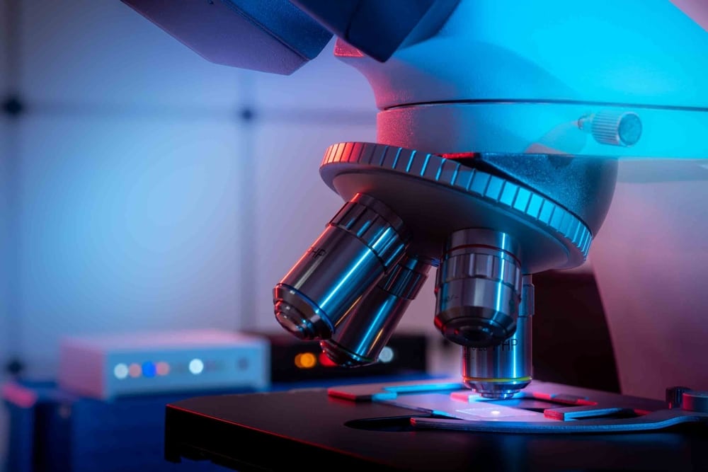
What is the actual appearance of a cell’s interior? Previously, conventional microscopes faced constraints regarding their ability to address this query effectively. Now, scientists from the Universities of Göttingen and Oxford, working in partnership with the University Medical Center Göttingen (UMG), have achieved the creation of a microscope with resolutions surpassing five nanometres (five billionths of a meter). This is approximately on par with dividing a hair into 10,000 separate strands. Their innovative approach was documented in Nature Photonics.
Numerous components within cells are so minuscule that standard microscopes only yield fragmented visuals. Their resolution commences at approximately 200 nanometres. For instance, human cells contain a type of framework composed of slender tubes that measure around seven nanometres in width. The synaptic cleft, describing the space between two nerve cells or between a nerve cell and a muscle cell, spans merely 10 to 50 nanometres – too diminutive for typical microscopes.
Advancement in Microscopy Technology
The newly created microscope, developed with assistance from researchers at the University of Göttingen, offers substantially richer data. It boasts a resolution exceeding five nanometres, allowing it to capture even the tiniest cellular structures. While it is challenging to envisage something so minuscule, if we were to equate one nanometre with one meter, it would be akin to comparing the diameter of a hazelnut to that of the Earth.
This class of microscope is referred to as a fluorescence microscope. Its operation hinges on “single-molecule localization microscopy”, where individual fluorescent molecules in a sample are activated and deactivated, followed by precise determination of their individual locations. Consequently, the overall structure of the sample can be reconstructed based on these molecules’ positions.
The existing methodology allows for resolutions around 10 to 20 nanometres. Professor Jörg Enderlein’s research team at the University of Göttingen’s Faculty of Physics has now managed to further enhance this resolution – utilizing a highly sensitive detector and specialized data analysis. This implies that even the minutest details of protein organization within the junction between two nerve cells can be depicted with great accuracy.
“This newly established technology marks a significant advancement in high-resolution microscopy. It not only provides resolutions within the single-digit nanometer realm but is also particularly economical and user-friendly compared to other techniques,” states Enderlein. The researchers have also created an open-source software package for data processing as part of their findings. This ensures that such microscopy techniques will be accessible to a broad spectrum of experts in the future.
Reference: “Doubling the resolution of fluorescence-lifetime single-molecule localization microscopy with image scanning microscopy” by Niels Radmacher, Oleksii Nevskyi, José Ignacio Gallea, Jan Christoph Thiele, Ingo Gregor, Silvio O. Rizzoli and Jörg Enderlein, 2 August 2024, Nature Photonics.
Image Source: luchschenF / Shutterstock


